


Cervical osteochondrosis or osteochondrosis of the cervical spine is a common disease of knowledge workers. Rapidly progressive disease. It is with cervical osteochondrosis, complicated by the development of disc herniation, that an increase in the incidence of early stroke is associated. For diagnosis, an MRI scan is required.
What is cervical osteochondrosis?
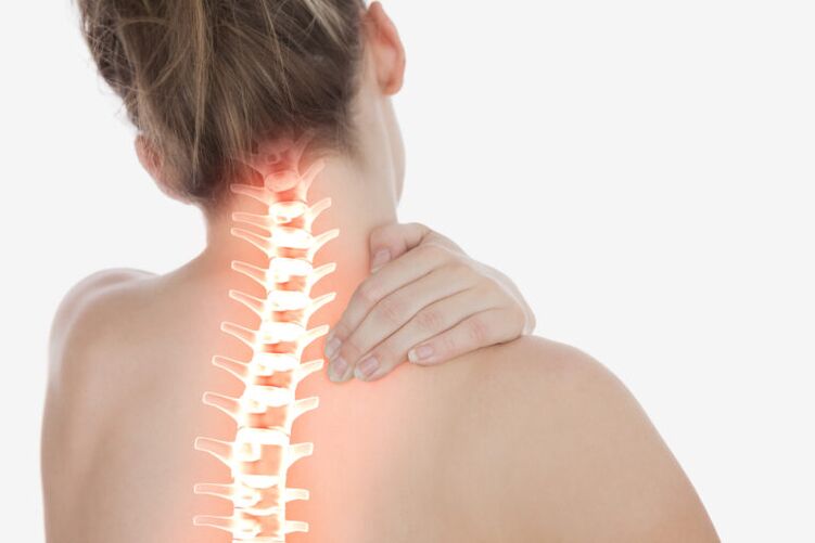
Cervical osteochondrosis is a common cause of neck pain, headaches, pressure surges, shoulder pain, numbness in the fingers, pain under the scapula. Currently, the frequency of cervical osteochondrosis has increased significantly, as the role of computers in our lives has grown.
However, a fall or injury can stimulate the onset of osteochondrosis, and degeneration (wear) of the intervertebral disc over time can lead to symptoms.
symptoms
In addition to moderate or mild pain, a feeling of stiffness in the neck and, in some cases, impaired mobility, many patients with cervical osteochondrosis feel numbness, tingling, and even weakness in the neck, arms, or shoulders due to chemicals. irritation and pinched nerves in the cervical spine.
For example, pinching a nerve root in the C6-C7 segment can cause weakness in the triceps, shoulder or forearm muscles, weakness in the wrist muscles, causing the hand to "hang", and changes in sensitivity in the middle finger.
Cervical osteochondrosis also often leads to the development of stenosis (narrowing) of the spinal canal and other progressive conditions, such as intervertebral hernias. How does this happen?
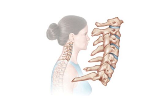
Osteochondrosis is nothing more than a degeneration of vertebral structures, caused, as a rule, by the natural aging of the body. With age, ligament thickening, bone growth formation on the vertebrae and other changes occur. When the spinal ligaments become thickened or bone growth appears, as well as for some other reason, there is less space for the spinal cord and nerves in the spinal canal. This condition is called stenosis, i. e. narrowing of the spinal canal. Severe narrowing of the spinal canal can cause compression of the nerve roots or even the spinal cord itself.
Intervertebral hernias are also, in most cases, the result of degeneration. The intervertebral disc serves as an absorber of frictional shocks between the vertebrae, thus preventing their destruction. Over time, the discs lose moisture and nutrients, become flattened, become more brittle and less elastic. As a result, cracks can form in the annulus, where part of the nucleus pulposus is squeezed out into the spinal canal. This condition is called an intervertebral hernia. If an intervertebral hernia compresses a nearby nerve root, pain syndrome and / or corresponding neurological symptoms occur.
Diagnostics
A successful diagnosis of cervical osteochondrosis begins with a doctor’s consultation. The doctor compiles the patient’s medical history and performs a physical examination to check for neck movements and sensitivity. During the examination, the patient may be asked to perform certain movements and report how pain symptoms change (increase or decrease).
If an examination shows that further testing is needed, your doctor may recommend radiographic tests such as radiography, magnetic resonance imaging (MRI), or computed tomography (CT). These diagnostic examinations, with varying degrees of reliability, can confirm the presence and localization of osteochondrosis, as well as identify other conditions (e. g. , calcification or arthritis) that may be the cause of a patient’s symptoms.
The best option for radiographic examination at present is MRI, because With the help of magnetic resonance imaging, it is possible to obtain high -quality detailed images of not only bone tissue, as in radiography, but also soft tissues of the spine, including muscles, ligaments, ducts, nerves andintervertebral disc. CT is usually prescribed if there are any contraindications to MRI, the main of which is the presence of metal structures or devices in the body (artificial joints, pacemakers, etc. ). The quality of a CT scan is lower than the quality of an MRI scan, but it can also show the condition of the soft tissues of the spine.
Treatment of cervical osteochondrosis
Conservative (non-surgical) treatment of osteochondrosis is always recommended as the primary strategy, and surgical intervention is only considered if complex conservative treatment for at least six months is fruitless or if pain and other symptoms significantly interfere with the patient’s daily routine. .
Methods used in the conservative treatment of cervical osteochondrosis may include:
- spinal traction. The no-load method of spinal traction, which has been used recently, allows to completely eliminate the complications of this method of treatment, without traction with a load is not possible. With increasing intervertebral distance, nutrition of all intervertebral discs improves, pain syndrome disappears.
- Rehabilitation gymnastics. . Rehabilitation gymnastics can increase the mobility of spinal segments. In the mobile vertebral segment, hernias and protrusions do not grow or form, because the intervertebral disc performs its function.
- massotherapy.
- drug therapy. Includes NSAIDs (non-steroidal anti-inflammatory drugs) and painkillers. In most cases, drug therapy has little or no temporary effect.
- cervical corset, orthopedic pillow. They can be recommended to stabilize the cervical spine and reduce stress on nerve roots after trauma and spinal fractures.
Surgical treatment of cervical osteochondrosis
If there is no significant relief after six months of conservative treatment and the daily routine becomes difficult for the patient, surgery may be considered. Typically, for cervical osteochondrosis, a procedure called spinal fusion is performed to paralyze the affected vertebral segment. This surgery involves removal of the intervertebral disc, decompression of the nerve root, and placing a bone or metal implant to maintain or create normal disc space and to stabilize the spinal segment.
As a rule, spinal fusion is performed on one vertebral segment, in rare cases, the question of performing surgery on two vertebral segments can be considered. However, patients should know that surgery to relieve neck pain symptoms is less likely to lead to a positive outcome than similar surgery to relieve pain in the arm with cervical osteochondrosis. Therefore, if neck pain is the main or only symptom, spinal fusion is only recommended as a last resort or if all conservative treatments have been tried and failed. If disc space cannot be identified as the most likely cause of neck pain, it is best to avoid surgery, although conservative treatment does not provide significant relief from the pain. In addition, do not forget that spinal surgery can be fraught with relatively serious consequences both in the operated area (local infection, rejection of implants, etc. ) and for the whole body (blood clots, allergic reactions to medications, etc. ). ). Therefore, before deciding on surgical treatment, it is necessary to discuss all the details of the operation directly with the surgeon who will perform it. It should also be noted that surgery on the cervical spine most often leads patients to vertebral deformities.
What is cervical osteochondrosis?
Official medicine interprets osteochondrosis as a degenerative-dystrophic lesion of the intervertebral disc.
From the part of the spine where this disc is located, a definition of the disease is also given.
Let us consider specifically the symptoms of cervical osteochondrosis, which accounts for almost 80% of all our back diseases.
The sad factor is that the disease affects the category of patients aged 30 to 50 years, that is, in the peak of their work capacity.
In young people, the disease acts as an independent disease, at an older age it is already a pathology that has developed against the background of other diseases of the joints.
How does the disease develop?
For any part of the spine, the gradual progression of the disease is characteristic. Cervical osteochondrosis does not go beyond this framework, so it deserves to be discussed in detail at each stage.
- In the early stages, there is a gradual destruction of the intervertebral disc. The fibrous annulus is located between them, where cracks appear, leading to a decrease in the elasticity and strength of the disc itself. They shrink and compress nerve roots.
- The second stage is the result of the first stage being untreated. The initial destruction of the disc spills into a chronic form, tissue compaction occurs, dislocation of the cervical vertebrae is observed. Falling head syndrome often develops at this stage.
- In the third stage, the sensation of pain increases, persistent headaches appear, the sensitivity of the upper limbs disappears, and the "lumbago" of the cervix is tortured. This is due to the fact that the fibrous ring at this stage is almost completely destroyed.
Often, there is a decrease in the pain sensation of third -degree cervical osteochondrosis. This occurs at this time when the cartilage tissue is gone and nothing is painful.

cause
Given the prevalence of osteochondrosis in general, doctors began to carefully study its causes. Many negative factors have been identified, but there is no definitive list. Here is what has been announced so far:
- sedentary lifestyle;
- all kinds of intoxication and infections;
- great physical activity;
- smoking;
- continuous weight lifting;
- stress and nervous tension;
- uncomfortable shoes or irregular feet that put unnecessary pressure on the spine;
- improper nutrition;
- frequent hypothermia and exposure to bad weather;
- age -related changes;
- spinal cord injuries;
- weak offspring;
- a sharp reluctance to train, if before that they had done so for a long time.
After familiarizing yourself with the causes, it becomes clear that cervical osteochondrosis can come at any age. And if at first the symptoms of osteochondrosis are insignificant and marked by rare attacks of pain, then over time it turns out it is impossible to turn the neck either, and it is difficult to tilt the head. And this is not the only danger of this disease.
What is the danger of the disease
Our necks are great workers. He was engaged all day, and his small vertebrae calmly withstood all the rounds and tilts of our heads. If the bones are removed, the nerves are pinched and the blood vessels are compressed, and the vertebral arteries, which are responsible for nourishing the brain, also pass through the cervical spine. Arteries are compressed, nerve roots are compressed and the inflammatory process begins.

What does this lead to? Spinal stroke, ischemia, intervertebral hernia - these are severe consequences of cervical osteochondrosis. We add here a general decrease in mobility and osteophyte formation. As a result, we experience disabilities that threaten complete immobility. With a disappointing prognosis, it is important to recognize the symptoms of cervical osteochondrosis quickly.
symptoms

The shortest way to identify osteochondrosis is a patient complaint. So what kind of sensation does a person discuss if his or her cervical vertebrae are destroyed? The picture of the disease looks like this:
- dizziness;
- Severe headache;
- "Flies" and colored spots in the eyes against the background of pain in the head;
- pain when turning, lifting weights;
- pain radiates to other organs (heart region, other organs).
Sometimes the signs of osteochondrosis can be listed among other diseases, but they should not be ignored, even if they are temporary in nature.
Diagnostics and treatment
As a continuation of the feelings expressed by the patient, the neurologist proceeded to diagnose the disease more accurately. A few years ago, only x-rays were in a doctor’s arsenal to recognize osteochondrosis. Computed tomography and magnetic resonance imaging are actively used today. They allow you to accurately determine the stage of the disease.
After evaluating the resulting image, a specialist vertebroneurologist prescribes the necessary treatment. The first thing doctors take is to relieve pain, then swelling and inflammation. To relieve pain, such anti-inflammatory drugs are used. As we recall, compression of the vertebral arteries disrupts the supply of the brain, which means that it needs to be repaired. This is done with the help of muscle relaxants.

Knowing that the symptoms and treatment of cervical osteochondrosis are related to the spine, massage and physiotherapy exercises must be included in the complex of health improvement measures. The massage is performed by a professional and by the patient himself. There are also special exercises aimed at developing the cervical vertebrae and restoring their mobility.